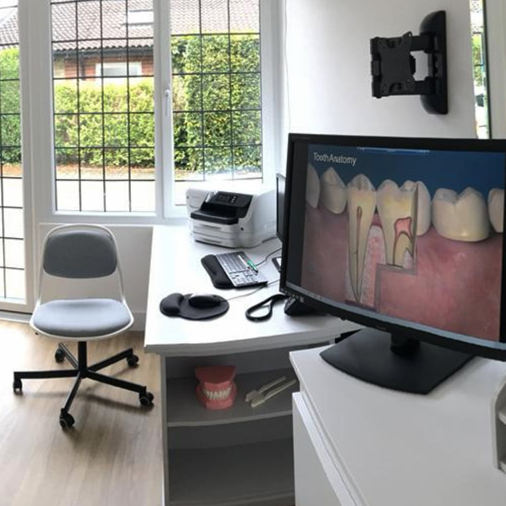
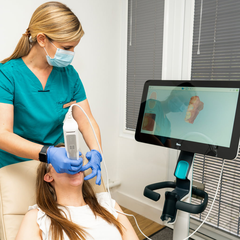
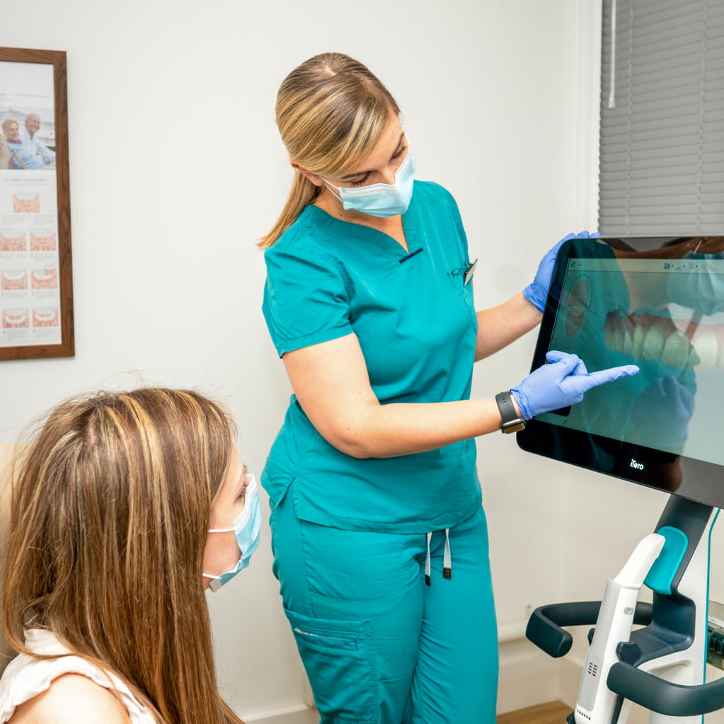
A dental X-ray is an X-ray imaging used to capture images of the teeth, gums, and jaws. Dental X-rays use low levels of ionising radiation to produce detailed images that allow dentists and dental specialists to evaluate oral health conditions that are not visible to the naked eye.
Dental X-rays are used to detect dental problems such as cavities, gum disease, dental abscesses, impacted teeth, and jawbone damage, among others. There are several types of dental X-rays, including bitewing X-rays, periapical X-rays, panoramic X-rays, and cone beam computed tomography (CBCT) scans, each of which is used to capture different views of the teeth and surrounding structures. Dental X-rays are safe, and the amount of radiation exposure is minimal, especially with digital X-ray technology.
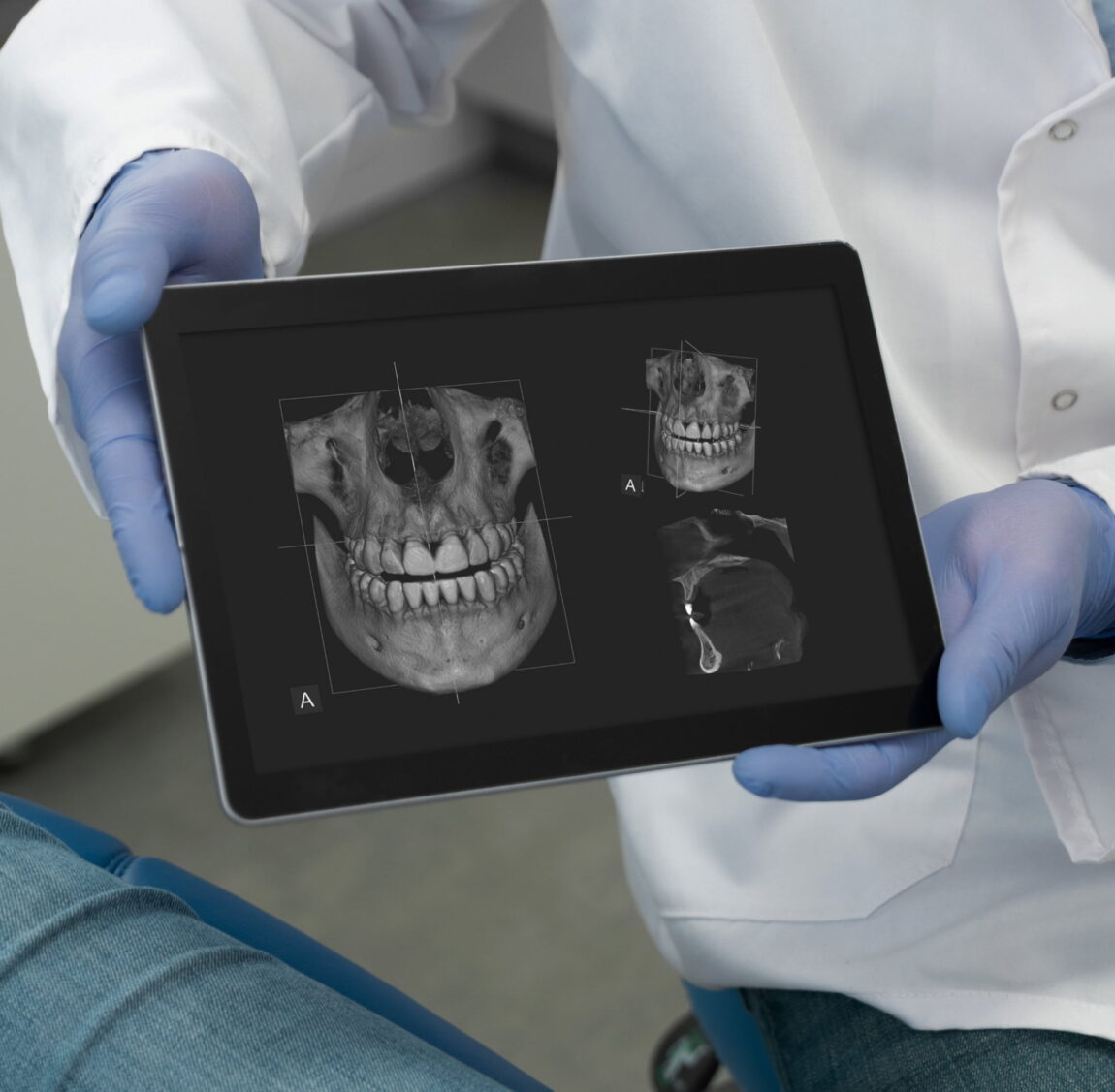
Cone Beam Computed Tomography (CBCT) is a specific form of imaging that uses a cone-shaped X-ray beam to produce detailed 3D images of teeth, jaws, and surrounding structures. CBCT has several dental applications, including:
Overall, CBCT is a valuable tool in dentistry, providing detailed 3D images that can help with diagnosis, treatment planning, and monitoring of dental conditions.
OPG is a type of dental X-ray that provides a panoramic view of the upper and lower jaws, teeth, and surrounding structures. OPG is a non-invasive and painless diagnostic tool that helps dentists and dental specialists to evaluate dental conditions, such as impacted teeth, fractures, infections, and bone loss, among others. The OPG machine rotates around the patient’s head, capturing a single, broad image of the mouth and teeth. OPGs are commonly used in routine dental check-ups, as well as before and after dental procedures, such as orthodontic treatment and dental implant placement.
Back to Dental Treatments


























 Dental Treatments

 FacialAesthetics

 Dental referrals


We offer innovative dental implant solutions to improve a smile.
Learn more
Innovative comfortable solutions for all your dentistry needs.
Learn more

 Instagram